becoming familiar with the microscope
© 2014 by KV5R. Rev. June 1, 2014.
Light Microscope Parts and Controls
A microscope is a relatively simple (yet precision) instrument, and is deliberately designed with hands-on controls for manipulating both the sample and the optical path. Learning the names and functions of the various parts and controls is an essential prerequisite to making an informed purchase, then properly using the microscope.
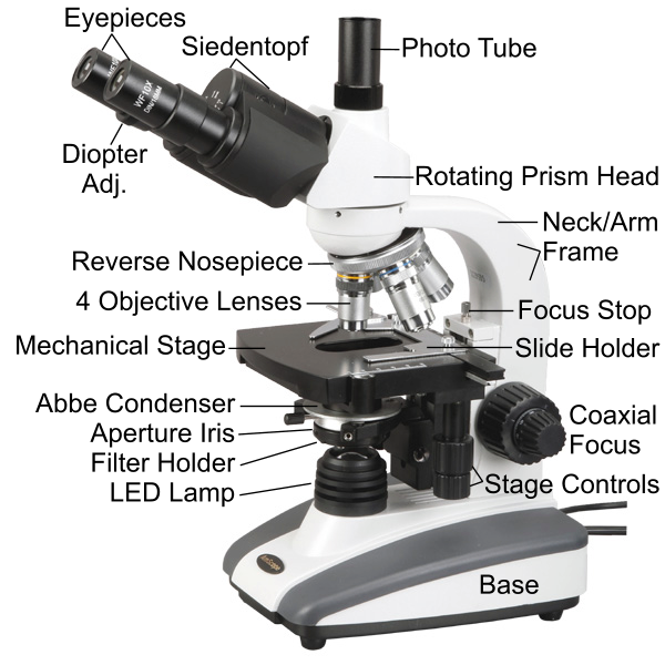
Trinocular microscope with Siedentopf head, photo tube, reverse nosepiece
mechanical stage, Abbe Condenser, and LED lamp (Amscope T360B
- Frame — this holds it all together as a useful machine. the upper part of the frame is called the neck or arm. The frame must be very rigid, as tiny relative movements are magnified in the image. Modern microscopes use cast aluminum frames and bases.
- Eyepiece (or ocular) lens — magnifies the image from the objective. The standard size for relatively inexpensive microscopes is 23mm diameter, 18mm exit pupil, 10 power. Some models also include other eyepiece powers like 16x or 20x. On models without a photo tube, the eyepiece lens may be replaced with a camera adapter for photo- and videomicrography. Eyepieces may be normal, wide-field, and/or extended-relief (longer viewing distance) for people with glasses.
- Ocular tube (monocular models) — holds the eyepiece (or camera/imager adapter) lens a fixed distance from the head prism, so that the eyepiece (or adapter) intercepts the rear focal plane of the objective. The conjugate distance from the objective flange to the eyepiece flange is a standard 160mm.
- Sliding Binocular head (binocular models) — holds two eyepieces for viewing with both eyes. The interpupillary distance is adjusted by sliding one side in or out. If your primary viewing will be on a computer screen (using a USB imager), you could buy a monocular microscope and save some money. Note that, unlike a stereo microscope, the binocular head on a biological microscope does not produce a 3D image; both eyes get the same image from the objective.
- Siedentopf Binocular head — a type of binocular head where the interpupillary distance is adjusted by rotating its halves around an off-center axis, like binoculars. Also called a “compensation-free” head, because, unlike the sliding type, the eyepieces do not need to be refocused when changing the interpupillary distance.
- Trinocular head — this is simply a sliding or Siedentopf binocular head with a third tube for a camera/imager adapter. This is more expensive but is a good choice if you will frequently switch between direct viewing and computer viewing, or you will be direct viewing and keep a still camera in the photo tube for quick photo acquisition.
- Rotating head — contains a 30 or 45-degree prism and a bearing that allows the angled eyepiece (or binocular assembly) to be turned in any direction. All modern microscopes (except some entry-level and pocket models) provide angled viewing, so the stage is kept level and wet samples may be observed. Note that microscopes with angled viewing are used from the front, with the user facing the stage, and not the back of the unit, as was the case with the old back-tilting straight-tube microscopes. It’s amusing to see how many “professionals” on TV shows are using modern microscopes backwards; indeed, many microscope ads picture them with the eyepiece assembly turned backwards. Traditions do die hard, if ever.
- Nosepiece turret — the rotating part that holds the 3 or 4 objective lenses, making lens changes easy. Some of the better models have a “reverse nosepiece,” which swing the unused objectives back under the frame arm, allowing easier access to the stage and specimens. They should all be made this way, as it is no more costly to do so. Having the objectives sticking out the front, in the way, is annoying, and is just a traditional hold-over from tilting microscopes that were used from the back.
- Objective lenses — the 3 or 4 lenses that directly observe and magnify the specimens on the slide. Various types are achromatic, plan-achromatic, plan-apochromatic, and phase-contrast. The quality of the objectives is the most important factor in the price and quality of the microscope.
- Stage — holds and manipulates the slide. In units with angled viewing, the stage (and thus the slide and sample) moves up and down for focusing, as opposed to the old straight-tube units where the stage was fixed and the entire optical assembly moved for focusing. Stages come in several types:
- Simple or plain — just a flat plane with a hole in it and leaf-spring clips to hold the slide down. The specimens in the sample are observed by pushing the slide around with the thumbs. Not suitable for higher magnifications. These appear in toy and entry-level educational microscopes.
- Simple, with add-on vernier slide holder — the leaf-spring clips of the simple stage are replaced with an add-on slide holder that uses finely-threaded shafts and knobs to move the slide around on the stage. They appear on mid-level educational microscopes, and will usually be called “mechanical” stages in the advertising. They should be called vernier slide holders, because that’s what they are. The stage itself does not move x-y, just the slide.
- Mechanical Stage — sometimes called a double-layer stage, this type has two levels with rack-and-pinion gears and linear bearings between them, and the slide holder is built right into the stage. They are controlled by two coaxial knobs that extend downward from the right-rear corner of the stage, quite near the focus. The top of the stage moves fore and aft only; left and right movements are performed by moving the slide holder sideways on the stage. Since moving around the sample is what we do the most, and the movements must be very fine and precise at high magnification, this type of stage is highly desirable and well worth a little extra cost. They are found on most advanced educational and all professional microscopes.
- Focusing Knobs, coarse and fine — these may be coaxial or separate, and they control the vertical position (z-axis) of the stage. Since they are the second-most used controls, they must be large, smooth-operating, on both sides, and without any annoying backlash. The focus knobs drive a pinion gear that drives a rack gear that attaches to the stage mount, which slides vertically in linear bearings within the frame. Coaxial focus uses a single rack-and-pinion, with a planetary reduction on the inner (fine) shaft. Seperate course and fine focus use two rack-and-pinion setups, each with its own set of knobs. The fine focus rack has a very limited travel, and you frequently run out of range and have to re-center it then move the course focus a little, then fine focus again. The coaxial type is much more desirable, since it uses only one rack and you never run out of fine focus range until you hit the focus limit stop.
- Focus Limit Stop — usually just a fine-thread screw with a lock-nut, located right behind the stage. It is set to keep the user from ramming the sample into the longer (40x and 100x) objective lenses and damaging or breaking the slide, the coverslip, or the objective. This is very important, as the working distance between the long objectives and the coverslip is well under 0.5mm. Note that the 40x and 100x objectives will usually have a spring-loaded lens assembly, allowing the lens to retract a few millimeters and reduce the potential for lens damage, but any touching of the lens to the coverslip may force it down against the specimen, immobilizing or mashing live critters between the slide and the slip. The stop screw is set (then locked) by carefully focusing the 100x lens on a tiny speck of dust or single cell on the slide. This is about about 0.10–0.15mm for most 100x objectives.
- Focus friction adjustment — usually a small lever or set-screw near the focus knob. It is adjusted tight enough to keep the stage from drifting downward during normal handling of the stage and sample. Models with coaxial focus usually do not have or need a focus friction, as the outer (coarse focus) shaft drives the inner (fine focus) shaft through a planetary bearing with a fairly high ratio and very thick grease, providing plenty of friction to keep the stage in position and maintain focus.
- Condenser — these focus and control the light that reaches the sample and the objective lens, fanning it out as a cone, just the right amount to fill the lowest element of the objective lens. Like stages, they come in several types:
- Simple — just a rotating disc with several aperture holes in it. Used on some toy models. These provide no adjustment for condenser focus, depth of field, or contrast.
- Fixed lens (in the stage hole) with rotating aperture disc — a little better than the above. Used on entry-level educational models.
- Abbe Condenser — a lens assembly that is suspended below the stage, having its own rack-and-pinion focus control. It contains 1, 2, or 3 lenses, an iris diaphragm (similar to a camera’s f-stop aperture), and a filter holder. The Abbe condenser assembly is raised or lowered, and the iris opening adjusted, to precisely control the light reaching the objective. The top of the top lens is flat, and is placed right below the slide (usually about 2mm). The position of the Abbe, and the iris, control the contrast and depth of field. They are usually supplied with advanced educational and professional microscopes, starting at around $200, and are highly desirable and well worth the extra cost. The lens system in the Abbe condenser may be uncorrected, achromatic, aplanatic, or even apochromatic in the very expensive models, but according to experts, the optical correction of the condenser is much less critical than the corrections in the objective lenses. The proper focus of the condenser may be determined by removing the eyepiece and looking directly into the tube, closing the iris diaphragm slightly (so the field of view is reduced to about 80%), and focusing the condenser until the edges of the iris leaves are in focus. The setting is different for each objective. The more expensive ones may have a top lens that can be swung out when using the low-power objectives.
- Variations of the Abbe condenser are made for darkfield, phase contrast, and other types for other purposes.
- Iris Control — a small lever that controls the opening of the iris, located near the bottom of the Abbe condenser assembly, just above the filter holder. Note that a condenser iris is not used to control brightness — that’s what the lamp dimmer is for — but is used to control the angle of the light cone that reaches the objective lens. In general, the effect on viewing is that an iris closed too far will produce multiple diffraction rings around objects in the sample, while one too far open will lose contrast and detail. Correctly adjusted, the specimens in the sample will be as sharp as possible without noticeable diffraction effects around them.
- Filter Holder — located just beneath the iris on the Abbe condenser assembly, it’s usually just a plastic ring that swings out. It holds not only filters, but anything you’d care to add to modify the light beam, such as darkfield stops, oblique stops, and multi-colored Rheinburg filters. Most microscopes have a 32mm filter holder. Some microscopes use a rotating filter turret below the condenser for fast-changing of various darkfield stops and/or phase contract annuli.
- Lamp Assembly — may be a simple mirror on very low-cost models (and most antiques), or may be an electric lamp with a dimmer control. The lamp itself may be tungsten, tungsten-halogen (longer life), fluorescent, or LED. By far the best, the modern white LED is cool, uses little power, lasts almost forever (≈50,000 hours), and best of all does not change color as it is dimmed. The power requirements of LED are so low that some LED microscopes are battery-powered for field use. Halogen lights use 20–50 watts, put out a lot of heat that will cook your specimens, accelerate evaporation, melt plastic filters, and typically last about 1500 hours. Worst of all, they change color, becoming reddish, when dimmed. Avoid halogen and go for the modern white LED—it really is worth the few extra dollars. Note that many modern microscopes are still being sold with halogen lamps, but they are gradually changing over to LED, making the halogen lamps obsolete.
- Base — the base should be stable and somewhat weighty, with vibration-isolating rubber feet. It usually houses the power supply, the lamp assembly, the fuse, the power switch, and dimmer control. The better desktop models have the power supply in the base instead of using a wall-wart. However, cordless microscopes
will have rechargeable batteries in the base and use a wall-wart for charging.
Light Microscope Standards and Features
Microscopes range from simple toys to complex instruments. The toys are fun in the field, but completely inadequate for the adult amateur microscopist. Educational microscopes, even at the lower-end, are considerably better than the toys, and at the higher end are fine for amateur microscopy. The advanced educational microscopes usually have many of the same features, and handle the same, as the lab-grade instruments, but at a much lower cost due to relatively inexpensive optics. What are the minimum features needed by the amateur microscopist? Here’s what I recommend:
- Standard sizes mean that you can upgrade or replace nearly every part of the microscope, if desired, and interchange with many different brands and types of optical components.
- Standard optical tube length: 160mm. This is the distance from the objective mounting flange to the top end of the eyepiece tube.
- Eyepiece diameter: 23mm. Some expensive models use 30 or 30.5mm.
- Objectives: DIN-standard (0.7965″×36TPI RMS threads, 45mm parfocal distance). DIN objectives are sometimes called 20mm (as in many ebay ads) to approximate the mounting thread diameter, but nowadays all objectives use the same RMS thread, and the only thing you have to consider (particularly when buying used) is the parfocal length, which will be 36mm JIS (Japan), or 45mm DIN (everywhere else), or perhaps something else for very old lenses (caveat emptor).
- Condenser holder diameter: 37mm.
- Filter holder: 32mm.
- Objectives: (4 achromats) 4x, 10x, 40x with spring-nose, and 100x oil immersion with spring-nose. At ≈$300 more, a set of plan (PL) objectives is highly desirable for photomicrography.
- Rotating head with 30 or 45 degree prism. Never consider the old straight-tube type that requires tilting the whole microscope back; you can’t comfortably view wet samples with them, and though collectible and nostalgic, are obsoleted by modern designs with level stage and angled prismatic viewing.
- Eyepieces: (2 wide-field) 10x and 20x. Get extended-relief eyepieces if you must wear glasses.
- Light: LED (1 watt or more) with dimmer
- Stage: mechanical double-layer with downward coaxial x-y controls
- Focus: coarse and fine, coaxial
- Condenser: Abbe-type with iris diaphragm and filter holder
- Digital USB imager (optional): 1.3 MP (1280x1024) is optimal for live viewing (about 15FPS) on USB-2, with its microscope adapter (≈$100)
A microscope with the above features costs about $200–400, will handle like a pro model, and is sufficient for all but the most demanding amateur microscopist, and even then, is easily upgraded with better optics and other accessories.
Note that you can increase its usefulness (and your enjoyment) with addition of simple home-made filters, darkfield stops, a couple 35mm linear polarizer slides, Rheinburg filters, etc. Half the fun is viewing your micro-specimens, the other half is playing with light itself!
Next, we’ll take a closer look at microscope types and quality, and the process of purchasing one.
—KV5R